Why Is The Brain Wrinkled?
Throughout human history, we have wanted to know the brain and each of its parts. We also wanted to know what the function of each of its regions is and know even its most unknown place. Today, we want to keep looking for answers, and the question we ask in this article is: why is the brain wrinkled?
Judging by the zoological scale, the relationship between neurons and learning ability is linear; the larger the surface of the brain, that is, the larger the cerebral cortex, the greater the learning. The human being has the maximum surface area and its brain is wrinkled thanks to the cerebral convolutions. In this way, it is possible to have a large surface in a small space.
Animals without convolutions are called lysencephalons and have little chance of learning, while animals called gyrebrains, endowed with convolutions, can learn more easily. The human being is the most gyencephalic being; its brain is more wrinkled than that of any other species, followed by the anthropoid apes.
Brain Anatomy: Differences Between Humans and Chimpanzees
In May 2009, the journal Scientific American published an article by Katherine S. Pollard, a biostatistician at the University of California, who developed software to compare the segments with the greatest difference between humans and chimpanzees. The sequence analysis showed that major difference comprises 118 nucleotides, which was called HAR1, ” h uman accelerated region ” (accelerated human region).
It appears that HAR1 is active in the human and other vertebrate brains. This region evolved very slowly in non-human vertebrates, as there are only two nucleotide changes between chicken and chimpanzee sequences, while the number of differences between humans is 18.
It has been shown in cultured cells that the HAR1 sequence is a modulator of gene expression, and was found to be active in a type of neuron involved in the development of the cerebral cortex. As a result, when cells where HAR1 is activated are damaged, the brain develops abnormally and the cerebral cortex does not look characteristically (with numerous sulci and lobes).
An anatomical feature of intelligence is not just the weight of the brain, but the fact that it is wrinkled, that is, the number of grooves and lobes it has.
How is it possible for the brain to be wrinkled?
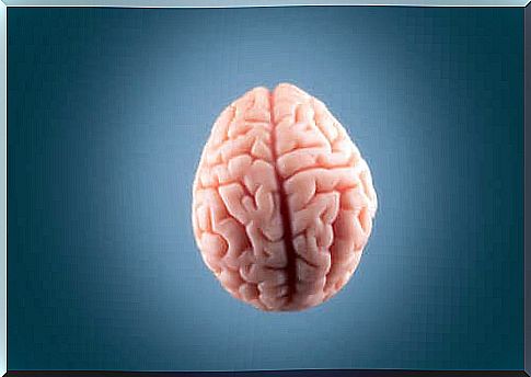
One of the most striking features of our brain is the incredible size of the cerebral cortex and its folds, visible as bumps and grooves on its outer surface.
As we mentioned, the majority of animals with a large brain cortex has a folded, while most of the animals with a small brain cortex has a smooth, wrinkle – free.
The cerebral cortex is a laminar tissue where neurons meet at the top. The bottom or inner part contains most of the connection between neurons and areas of the brain.
In large brains, this layer of neural tissue that covers the outside of the brain is disproportionately larger than the deep brain structures it covers. In Instead of adopting a balloon shape, bending it by minimizing the total volume of the brain and cranium.
Victor Borrel and his team have been researching this area for several years and have shown that basal radial glia (bRG), regulated by intrinsic and extrinsic factors, plays a key role in the tangential expansion of the cerebral cortex.
Thus, the BRG is an essential requirement, but not sufficient to generate a cortex with curved, it provides new processes by which the radial neurons migrate radially, tangentially dispersing and expanding the cerebral cortex.
What happens when the brain is not properly wrinkled?
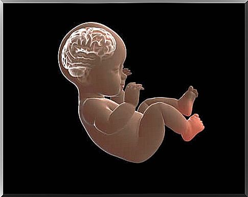
Cortical folding occurs during embryonic development. The wrinkled form of the brain develops around the twentieth week of pregnancy and is complete when the child is one and a half years old.
This is important for optimizing the brain’s functional organization and connections. Furthermore, it allows you to adjust a large cortex to a cranial volume limited by its size.
Some of the most common pathologies are polymicrogyria (formation of several small turns) and periventricular nodular heterotopia. In the latter, neurons accumulate ectopically in the vicinity of the telencephalic ventricles, forming nodules that act as an epileptic focus.